Sunday, March 13, 2016

Intracranial Hypertension Diet

Intracranial Hypertension Diet

Intracranial hypertension or pseudotumor cerebri occurs when the pressure within the skull, known as intracranial pressure, increases for no evident reason. The symptoms are similar to those of a brain tumor. It occurs most commonly in obese women during their childbearing years but it may also affect children and adults. Intracranial hypertension causes a buildup of cerebrospinal fluid. This in turn causes swelling in the brain.

The blood volume in the blood vessels surrounding the brain also increases. While most people with this condition can continue leading normal lives, others need to take certain precautions such as avoiding certain sporting activities and keeping a check on their diet.

Foods to include


Foods that are good for intracranial hypertension include:
  • Fruits – Fruits contain fiber and vitamins and form an important part of any diet for weight loss. Keep a bowl of fruit handy and eat at least 5 helpings every day. They make a wonderful snack between meals and help to keep the hunger pangs at bay.
  • Lean meat – poultry and fish have low fat content and can help to keep the calorie count low while supplying your body with the proteins it needs.
  • Low fat dairy products
  • Olive oil, canola oil and other sources of monounsaturated fats will help keep cholesterol levels under control.
  • Oats
  • Whole grain bread
In addition to these foods for intracranial hypertension, it is essential to maintain a healthy lifestyle and exercise regularly.

Foods to Avoid


Certain foods lead to an increase in intracranial pressure and should be limited or avoided altogether. Your doctor may suggest certain changes to your regular eating habits. Some of these restrictions include:
  • A limit your intake of salt. Salt causes water retention within the body and causes an increase in blood pressure.
  • A limit your intake of fats. Excessive levels of fat in the diet hinder weight loss that is advisable for this condition. Avoid red meat and processed meats like pork, bacon and beef.
  • Foods that are rich in vitamin A and tyramine may have an adverse impact on intracranial hypertension. Tyramine is a compound formed during the breakdown of tyrosine, an amino acid and can cause blood vessels to dilate. For this reason, foods rich in tyramine should be avoided. These include preserved, dried or aged foods such as pickles, pickled foods, salami, pepperoni, sauerkraut, olives, fermented soy products, nuts, aged cheese, beer and wine. Foods with high levels of vitamin A should also be avoided. These include liver, carrots, tomatoes, sweet potatoes and green leafy vegetables.
  • Alcohol – If you must drink make sure you consume enough water to prevent dehydration.
  • Coffee – This includes drinks containing caffeine.

Intracranial hypertension and obesity


The exact relationship between intracranial hypertension and weight is not known. Many people with intracranial hypertension however, report a decrease in symptoms such as papilledema, with a reduction in weight, while in some instances, remissions have been known to occur. For this reason, reduction in weight is very important for those who are overweight and have intracranial hypertension. A correct diet for intracranial hypertension can help people lose weight and may lead to a decrease in symptoms associated with this condition. Studies show that a low energy diet is very effective in losing excess weight and reducing the symptoms of intracranial hypertension.
There are also certain activities you need to avoid if you have intracranial hypertension. . These include activities such as somersaults, bungee jumping, judo, rugby, high g-force rides at amusement parks, and twisting and stretching exercises. Avoid excessive exposure to the sun as intracranial hypertension can cause problems with body temperature control. You should also quit smoking. Intracranial hypertension may also affect your ability to judge distances and speed.
Additional intracranial hypertension info
Symptoms
Symptoms of intracranial hypertension include:
  • Blurred vision
  • Episodes of blindness in one or both the eyes that last for a few seconds
  • Headaches originating behind the eyes which get aggravated with eye movement
  • Ringing in the ears
  • Difficulty in seeing to one side
  • Double vision
  • Seeing light flashes
  • Pain in the neck, shoulder or back
Causes
In most cases the causes of intracranial hypertension remain unknown. It may be linked to an excess of cerebrospinal fluid within the skull as well as to a narrowing of the transverse sinuses. In the latter case it is uncertain whether the narrowing of the transverse sinuses is a caused by or an effect of the condition.
Treatment
Treatment of intracranial hypertension begins with medications that control the symptoms. These include:
  • Glaucoma drugs – Acetazolamide (Diamox) is the drug of choice. It cuts the production of cerebrospinal fluid by at least 50%
  • Diuretics – If acetazolamide alone proves ineffective, it may be combined with a diuretic such as furosemide which helps reduce liquid retention
  • Migraine medications – These are sometimes given to treat the severe headaches caused by intracranial hypertension.
Surgery is sometimes necessary to reduce intracranial pressure or pressure on the optic nerve. Surgical procedures include:
  • Optic nerve fenestration – A cut is made in the membrane surrounding the optic nerve allowing excess cerebrospinal fluid to drain away.
  • Spinal fluid shunt – A thin long tube or shunt is inserted into the brain or lower spine which helps drain away excess cerebrospinal fluid.
Stages
There are three stages intracranial hypertension. In stage 1, the intracranial pressure increase is minimal. Stage 2 is characterized by a drastic increase in intracranial pressure. In stage 3 the increases is so severe that it causes a reduction in cerebral flow, eventually leading to ischemia and brain infarction.

References

Benign Intracranial Hypertension Symptoms

Benign Intracranial Hypertension Symptoms

Overview

When the pressure inside of the skull becomes elevated for no clear reason, it leads to a condition called benign, or idiopathic, intracranial hypertension. Also referred to as pseudotumor cerebri, benign intracranial hypertension symptoms can falsely mimic those caused by a brain tumor. Though problems caused by the condition are often reversible, it is vital that you seek treatment right away to prevent permanent and serious complications from occurring.

Head Symptoms

According to the UCLA Health System, a headache is the most common symptom of benign intracranial hypertension. Reported in approximately 94 percent of all patients, headaches can be extremely severe or fluctuate in intensity, and they are often worse in the morning hours. Almost one-third of patients will experience a feeling of lightheadedness or dizziness, as well as nausea, a symptom that may be brought on by the headache.

The National Institutes of Health advises that babies with benign intracranial hypertension may also have a noticeably larger head. Very young infants whose skull sutures have not yet closed may demonstrate a bulging around the fontanelle, or soft spot.

Papilledema and Vision Changes

Adverse effects on vision are also very common symptoms of benign intracranial hypertension, found in roughly 48 percent of patients, according to the UCLA Health System. Affected individuals may notice intermittent periods of blurred or distorted vision, usually starting with their peripheral vision. Diplopia, or double vision, is also quite common. According to the Merck Manuals Online Medical Library, these minor changes in vision often go unnoticed until intracranial hypertension has progressed significantly. Permanent partial or complete vision loss is possible in some cases.

Papilledema, a swelling of the optic nerve of the eye, is also a common symptom caused by increased pressure inside of the skull. According to Merck Manuals, papilledema usually occurs in both eyes, although some people may develop it in one eye only or not at all. The condition sometimes causes momentary changes in vision, but usually must be detected through an eye exam conducted by an ophthalmologist.

Tinnitus

Benign intracranial hypertension may also cause symptoms of tinnitus, or a ringing in the ears. In addition to a ringing sound, some people may hear a buzzing, hissing, whirring or roaring sound. According to Merck Manuals, pulsatile tinnitus--a rhythmic noise matching the beat of the pulse or heart that is caused by a change in the rate or pressure of blood flow in the vessels around the ears--is especially common among those with pseudotumor cerebri.

Pseudotumor Cerebri

What Is Pseudotumor Cerebri?

Pseudotumor cerebri is a condition in which the pressure around your brain increases, causing headaches and vision problems. The name means “false brain tumor” because its symptoms are similar to those caused by brain tumors. It’s also known as idiopathic intracranial hypertension. This condition is treatable, but it can return in some cases.

What Causes Pseudotumor Cerebri?

Causes Icon
The exact cause of this condition is unknown, but it may be associated with having too much cerebrospinal fluid in your skull. This fluid, which protects your brain and spinal cord, is normally absorbed into your bloodstream. Pseudotumor cerebri may occur when this fluid isn’t fully absorbed, which causes it to build up. This leads to increased pressure in your skull.

What Are the Risk Factors for Pseudotumor Cerebri?

Risk Factors

Obesity

Obesity is one of the leading factors that can increase your risk of developing pseudotumor cerebri. According to the Mayo Clinic, the risk is almost 20 times higher in obese women who are under 44 years old than in the general population.

Medications

Certain medications may make you more susceptible to this condition. These include:
  • birth control pills
  • excessive amounts of vitamin A
  • tetracycline
  • steroids (when you stop using them)

Other Health Conditions

Health conditions associated with pseudotumor cerebri include:
  • kidney disease
  • sleep apnea, which is abnormal breathing during sleep marked by phases of paused breathing
  • Addison’s disease, which is a disorder in which your adrenal glands don’t produce enough hormones
  • Lyme disease, which is a chronic flu-like disease caused by a bacterium carried by ticks

A Birth Defect

Stenosis is a condition that causes narrowing of the blood vessels in your brain. It may make you more likely to develop pseudotumor cerebri. The narrowed veins make it more difficult for fluid to move through your brain.

What Are the Symptoms of Pseudotumor Cerebri?

Symptoms Icon

Headaches

A common symptom of this condition is a dull headache that starts behind your eyes. These headaches can become worse at night, when you move your eyes, or when you first wake up.

Vision Problems

You may also have vision problems, such as seeing flashes of light or having brief episodes of blindness or blurred vision. These problems can become worse as the pressure keeps increasing. This can lead to double vision or permanent vision loss.

Other Symptoms

Other symptoms include:
  • ringing in your ears
  • pain in your neck, back, or shoulders
  • nausea
  • vomiting
  • dizziness
  • neck or back pain
  • d

How Is Pseudotumor Cerebri Diagnosed?

Diagnosis Icon

Eye Exam

Your doctor will check for papilledema, which is swelling of the optic nerve at the back of your eye. Your vision will also be tested to see if you have abnormal blind spots.

Imaging Tests

Your doctor may perform a CT or MRI scan of your brain to look for signs of spinal fluid pressure. These scans can also be used to check for other conditions that could be causing your symptoms, such as tumors or blood clots.
A CT scan, combines several X-rays to make a cross-sectional image of your brain. An MRI scan uses radio waves and magnets to produce a highly detailed image of your brain.

Spinal Tap

Your doctor may also perform a spinal tap, or lumbar puncture, to measure the pressure of your spinal fluid. This involves placing a needle between two bones, or vertebrae, in your back and drawing a fluid sample for testing.

What Are the Treatments for Pseudotumor Cerebri?

Treatment Icon

Medications

Medications can help control or reduce the symptoms of pseudotumor cerebri. Your doctor might prescribe the following:
  • Migraine medications can provide headache relief. These can include triptans like sumatriptan (Imitrex) and naratriptan (Amerge)
  • Glaucoma drugs, such as acetazolamide (Diamox), cause your brain to produce less cerebrospinal fluid. These drugs can cause fatigue, kidney stones, nausea, and a tingling sensation in your mouth, toes, or fingers.
  • Diuretics, such as furosemide (Lasix), make you urinate more often. This causes you to retain less fluid in your body, which helps ease the pressure in your skull. These may be used in combination with glaucoma drugs to make them more effective.

Surgery

Your doctor may recommend surgery if your vision becomes worse or if they need to drain excess cerebrospinal fluid.

Optic Nerve Sheath Fenestration

Optic nerve sheath fenestration involves cutting the membrane around your optic nerve to let extra fluid out. According to the Mayo Clinic, it’s successful at relieving symptoms more than 85 percent of the time.

Spinal Fluid Shunt Placement

A spinal fluid shunt procedure involves placing a thin tube in your brain or lower spine to drain extra fluid. This procedure is usually done only in severe cases. According to the Mayo Clinic, it has a success rate of more than 80 percent.

Other Forms of Treatment

Other treatment methods include losing weight and having multiple spinal taps performed to relieve pressure.

Post-Treatment Outlook

Icon Outlook
You’ll need to see your eye doctor regularly to have your vision checked once the pseudotumor cerebri is gone. Your eye doctor will watch you closely to make sure that you don’t continue to have vision changes that could result in permanent vision loss.
You should also let your primary care doctor know if you start having symptoms of this condition again.

Can Pseudotumor Cerebri Be Prevented?

Prevention Icon
Gaining weight puts you at a higher risk of having a pseudotumor cerebri. You can help prevent this condition by losing excess body weight and keeping it off. Switching to a healthy diet and getting regular exercise can help you drop the extra weight.
Your diet should include plenty of fruits, vegetables, and whole grains. You should also choose lean meats and dairy products that are low in fat. Limit or avoid eating foods that are high in:
  • added sugars
  • saturated fat
  • trans fat
  • sodium
Adopt a regular exercise routine, which can be as simple as walking. You can follow a more vigorous workout routine if your doctor says it’s safe to do so.


Wednesday, March 2, 2016

Idiopathic Intracranial Hypertension

http://webeye.ophth.uiowa.edu/eyeforum/cases/99-Pseudotumor-Cerebri.htm

Idiopathic Intracranial Hypertension

(Pseudotumor Cerebri)

Leslie Pham, MDMichael Wall, MD

December 17, 2009, updated February 2, 2010

Chief Complaint: Headaches and transient double vision for 6 months

History of Present Illness:

A 31-year-old obese female presented to her eye doctor with a history of headaches and transient visual obscurations for six months. She also reported a ringing sound in her ears for the past year that was synchronous with her pulse. On exam, she was noted to have bilateral optic disc edema. An MRI scan was performed and was normal except for an empty sella. The MR venogram was normal with no evidence of sinus thrombosis. She was diagnosed with idiopathic intracranial hypertension and referred to a neurologist for further workup and management. The neurologist performed a lumbar puncture which showed an elevated opening pressure of 50 cm H2O. The patient was started on acetazolamide (Diamox®) 500 mg twice a day and encouraged to lose weight.

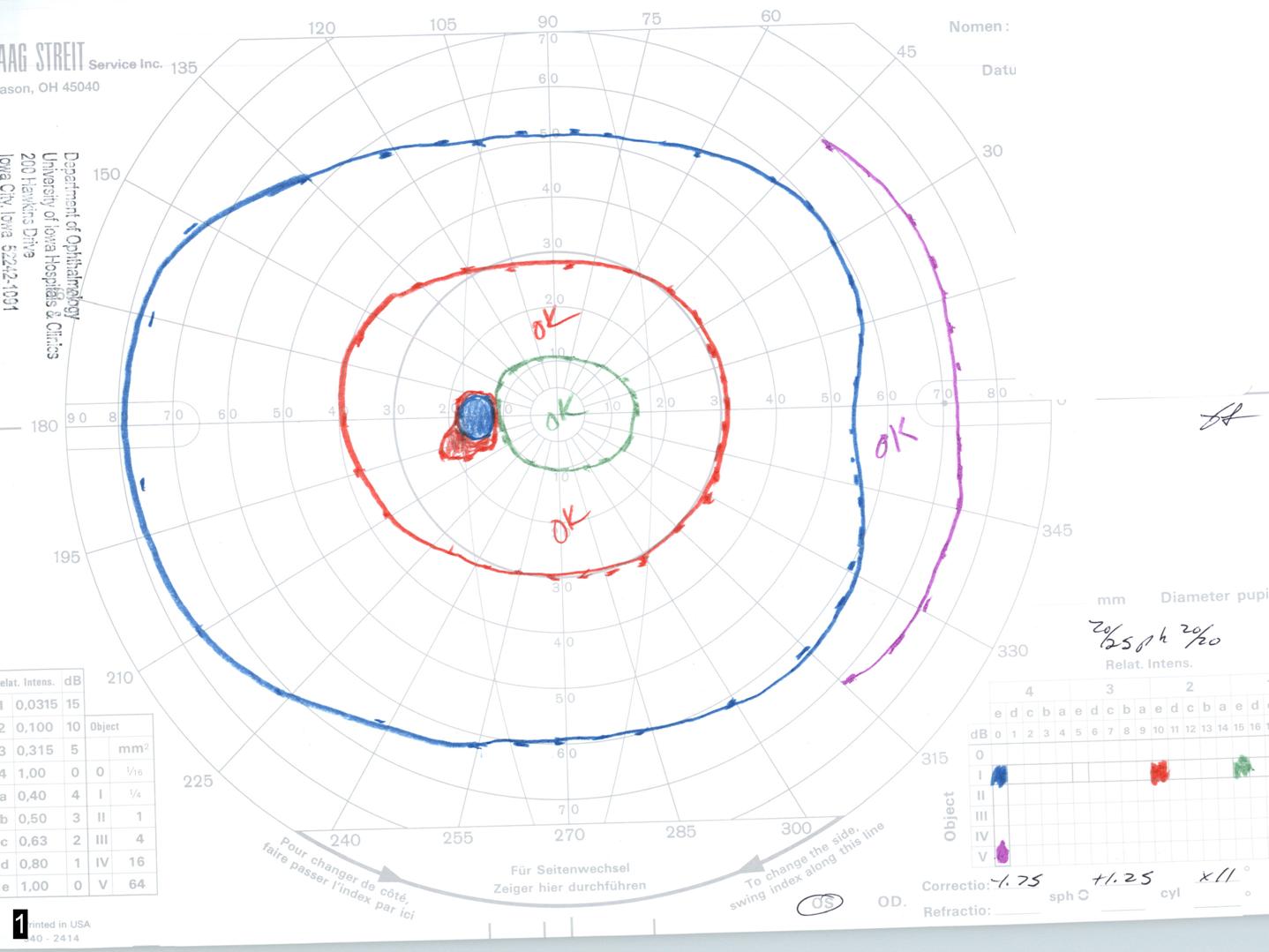
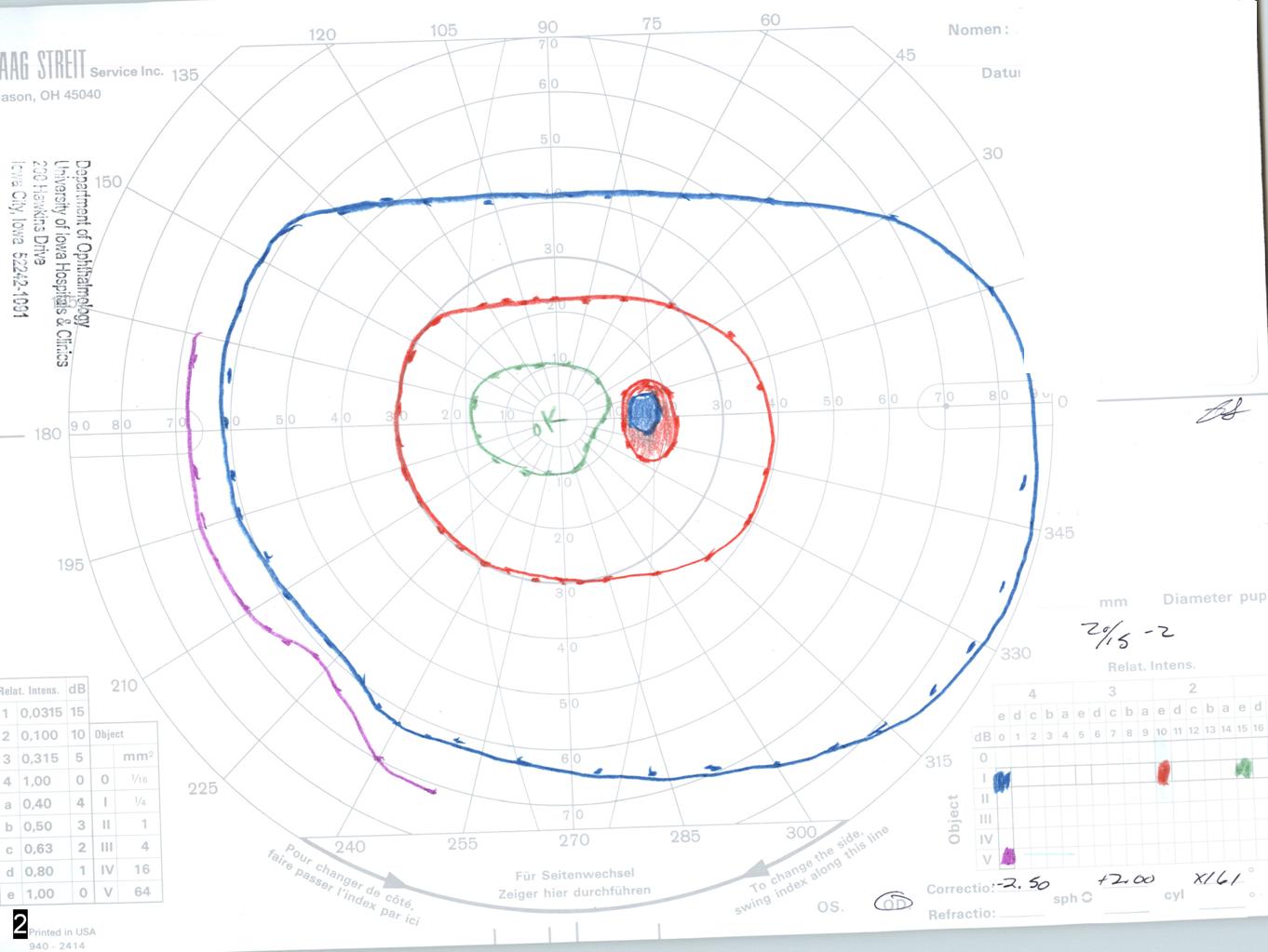
The patient continued to have severe headaches after the lumbar puncture. One month later, she returned to her eye doctor for visual field testing. Humphrey perimetry was performed, and showed a mean deviation of -20.85 in the right eye with enlargement of the blind spot and a dense nasal field defect. In the left eye, the mean deviation was -20.35 and also showed enlargement of the blind spot and a dense nasal field defect. A repeat lumbar puncture was performed, which again showed an elevated opening pressure of 50-56 cm H2O. Her Diamox dosage was increased to 500 mg three times daily and she was subsequently was referred to the Neuro-ophthalmology Service at UIHC for further evaluation and management.

Past Ocular History: Prior eye examination at UIHC in 2000 was normal.

Past Medical History: Obesity and Depression.

Medications: Acetazolamide (Diamox®) 1500 mg daily, citalopram hydrobromide (Celexa®)

Family History: Non-contributory

Social History: Non-smoker, denies alcohol use

Review of Systems: She reports significant weight fluctuations over the last several months.

Physical Exam:

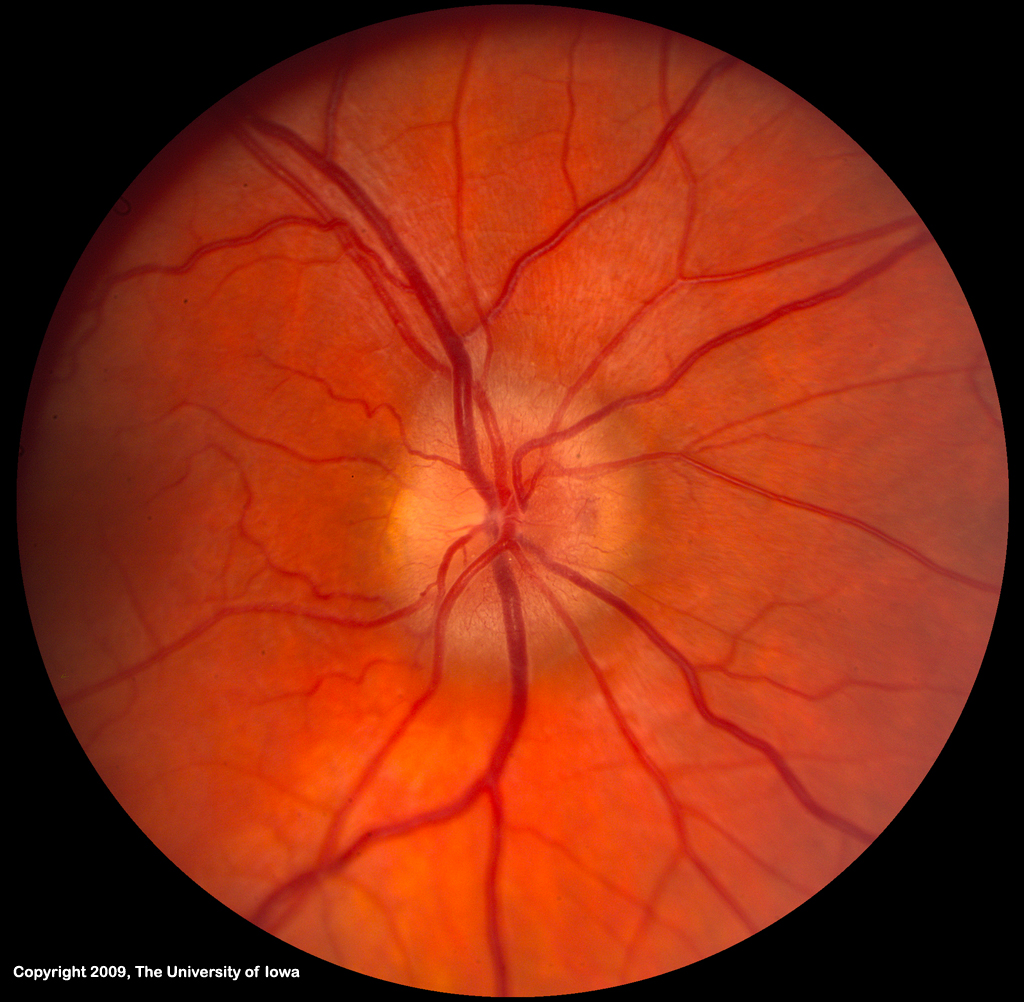
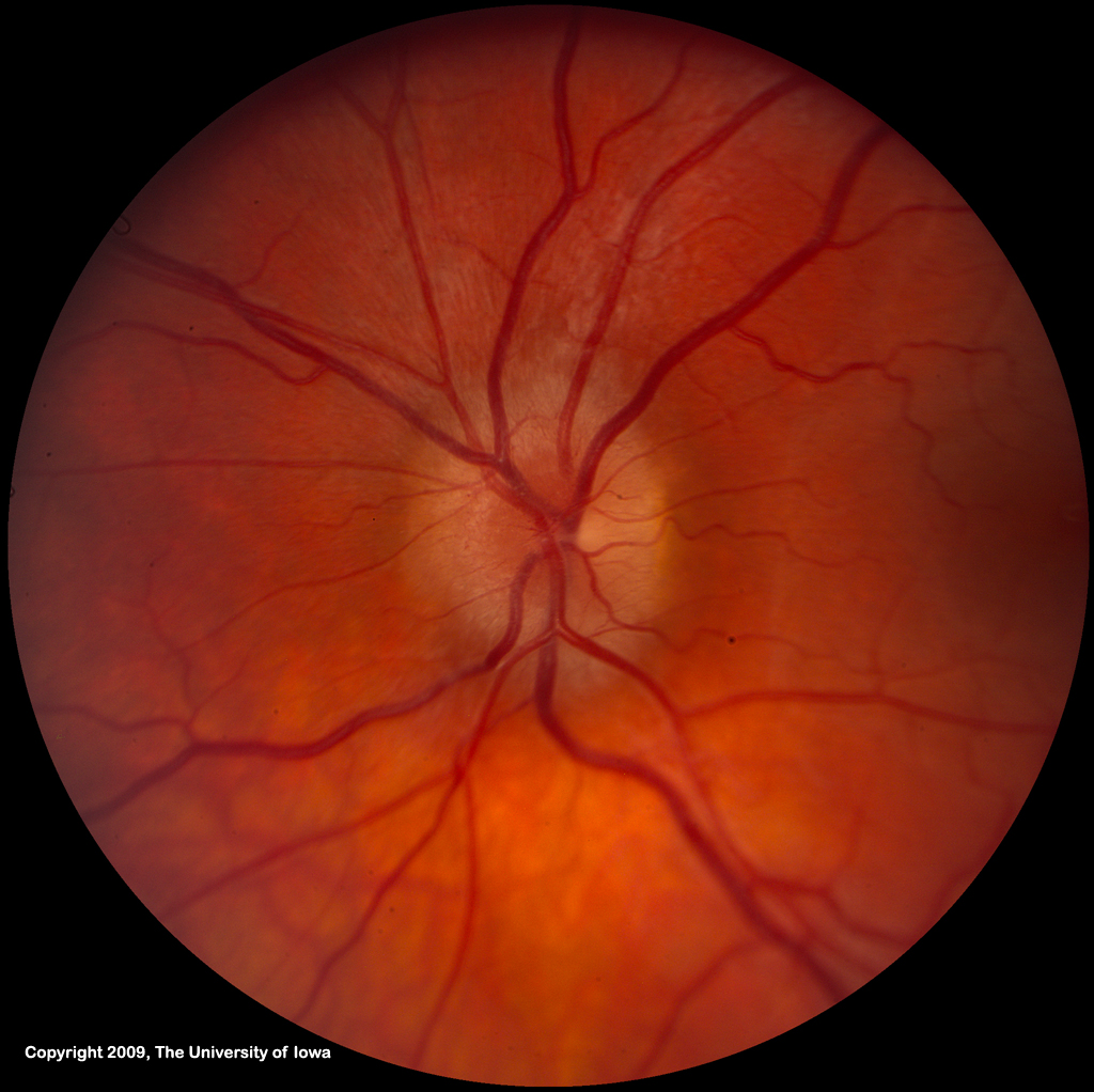
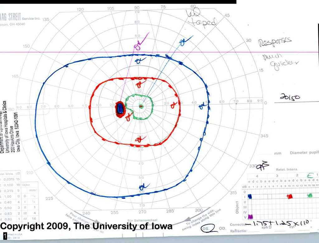
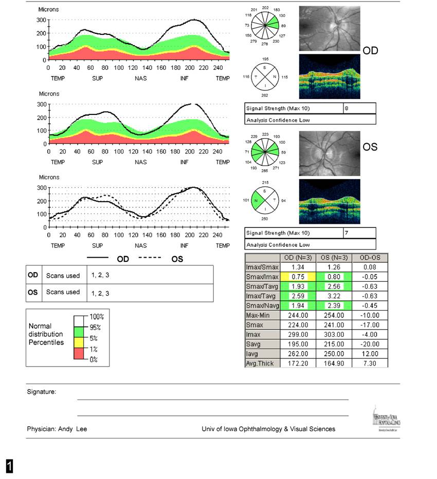
Visual AcuityOD-- 20/20OS-- 20/50Extraocular motility: full OU, without diplopiaPupils: 5 mm in the dark and 2 mm in the light; no relative afferent pupillary defect.Intra-ocular pressure: 11 mmHg OD, 16 mmHg OSColor vision testing normal in each eye.She had normal cranial nerve V and VII function bilaterally.External and slit lamp exam normalThe patient had grade two papilledema. The macula, vessels and periphery in each eye were normal. (see figure 1)Figure 1. Bilateral optic disc edema at presentation, (12/07) Goldmann perimetry was performed at her initial visit (click on either image to enlarge)Figure 2. Goldmann visual field at presentation (12/07) -- (click on either image to enlarge)The right eye field shows a markedly enlarged blind spot and some nasal constriction. The left eye field shoes a small paracentral scotoma to the 11e and 12e isopter, but full peripheral field. The patient reported having transient visual obscurations during testing of her left eye.Figure 3. Optical coherence topography at presentation (12/07) demonstrates marked elevation of the retinal nerve fiber layer OU. (click on image for larger view)

Course

The patient's dose of Diamox was increased to 2 gm daily, given that she had papilledema, persistent headaches, transient visual obscurations and pulse synchronous tinnitus. When she returned two weeks later, she felt better. She had lost 5.5 pounds and her headaches had resolved. Her visual acuity was 20/20 OD and 20/25 OS and ophthalmoscopy showed residual grade I-II disc edema. Her Goldmann visual field had improved. She was kept on the same dose and returned for follow up, 2 months later.

At her follow up appointment she reported only three headaches since her last exam. She had lost an additional 4.5 pounds for a total of 10 pounds of weight loss. Goldmann visual field showed a slight enlargement of the blind spot on the left greater than the right. The rest of the field was full. Her visual acuity was 20/15 OD and 20/25 OS and ophthalmoscopy showed grade I optic disc edema. (see figures 3 and 4)

She has since been doing well and her Diamox was tapered off without further complications. She continues to be followed by the neuro-ophthalmology clinic.

Figure 4. Optic disc edema improved, (3/08) -- (click on either image to enlarge)Figure 5. Goldmann visual fields are much improved from prior, with normalization of blind spot size and disappearance of the paracentral scotoma in the right eye (3/08) -- (click on either image to enlarge)

Discussion

Idiopathic Intracranial Hypertension (IIH) is a condition in which the cerebrospinal fluid (CSF) is under high pressure in the absence of an intracranial mass, venous sinus thrombosis or other primary cause (see differential diagnosis). It is also known as pseudotumor cerebri because patients present with signs and symptoms of a brain tumor without a brain tumor being present.

Pathophysiology

In IIH, there is no structural obstruction to the circulation of CSF. It is believed that the problem lies in defective reabsorption of CSF either at the level of the arachnoid granulations or along cranial and spinal nerve root sheaths. There is no evidence for excessive CSF production.

There is a female preponderance for this disease, and the peak age of onset is in the third decade of life. It is common for affected patients to be obese, and weight loss is an important part of treatment. IIH symptoms often start or worsen during a period of weight gain and resolve with 5-10% total weight loss. The disease is rare in thin men. This has led some researchers to look for an association with hormonal changes within the body, but to date no consistent associations have been found. The disease has been associated with withdrawal from steroids, and with the use of exogenous substances like vitamin A, tetracyclines, and oral contraceptives.

Although no associated conditions besides recent weight gain are usually found, many conditions have been linked to high intracranial pressure. Any disorder that blocks the flow of spinal fluid between the brain and the jugular venous system can cause raised pressure. For example, scarring of the arachnoid granulations by meningitis can cause raised intracranial pressure. Similarly, blood clots in the veins draining the brain can cause increased intracranial pressure. These conditions can mimic IIH.

Symptoms and Signs

IIH presents with symptoms and signs of elevated intracranial pressure. Headache is the most common symptom. The headache may be aggravated by changes in position. There may be a ringing or a "whooshing" sound in the ear synchronous with the patients pulse. A patient may experience double vision. Momentary loss of vision known as "transient obscurations of vision" are common; they may occur in one or both eyes and usually last seconds but rarely longer than one minute. Other visual symptoms are dimming of vision, or loss of peripheral vision in one or both eyes. The visual loss may be mild to severe. If severe and untreated, the disease can result in permanent vision loss.

A prospective study conducted by Wall and George demonstrated the following incidence of symptoms

headache (94%)transient visual obscurations or blurring (68%)pulse synchronous tinnitus or "whooshing noise" in the ear (58%)pain behind the eye (44%)double vision (38%)visual loss (30%)

Over 90% of patients in this study had visual loss documented by perimetry (Wall and George, 1991).

On examination, papilledema (bilateral optic disc edema) is nearly always present. This may be accompanied by a serous retinal detachment and choroidal folds if the intracranial pressure is excessively high. (seeimages of Grades I-V papilledema)

Images of Grades I-V papilledema (click on image for details)

Diagnosis

First, imaging studies of the patient's head (via magnetic resonance imaging or computed tomography) must be normal except for the signs of raised intracranial pressure (empty sella, flattened globes, collapsed lateral sinus). Next, the cerebrospinal fluid pressure is measured by a lumbar puncture and the pressure must be elevated. The lumbar puncture should be performed with the patient in a supine position for most accurate measurement of the intracranial pressure. Laboratory studies on the cerebrospinal fluid must be normal. All of the above criteria (the Modified Dandy Criteria, see table) must be met in order to diagnose IIH.

Clinical Definition of IIH" (Modified Dandy Criteria)Signs and symptoms of increased intracranial pressureNo localizing neurologic findings (Cranial nerve VI palsies are allowed)Normal neuroimaging (with the exception of an empty sella)Opening pressure of lumbar puncture of greater than 250 mm water, with normal CSFNo other cause of increased intracranial pressure present

Treatment

A variety of treatments for IIH have been proposed and there is much anecdotal evidence for these treatments. There is no evidence-based data currently available but an NIH sponsored clinical treatment trial is beginning in 2009. Patients with no visual loss or mild visual loss are treated medically.

Weight loss is an important part of treatment and is always recommended. Most patients improve after losing 5-10% of their total body weight. It appears that in addition to weight reduction, sodium restriction is also a useful adjunct. We recommend that patients lose about one pound a week for several months and then maintain the weight loss. The mechanism of the beneficial effect of weight loss on this disease is unclear but there are parallels with the beneficial effect of weight loss and sodium restriction for treatment of essential hypertension.

For patients requiring medical therapy, acetazolamide (Diamox®) is the most commonly used medication. It is relatively safe but nearly all patients experience the side effect of paresthesias, or numbness and tingling in the extremities. Patients also report an alteration in taste such that carbonated soft drinks taste metallic. Less commonly, kidney stones can occur. Another diuretic commonly used that appears to be effective in some patients is furosemide (Lasix®). Topiramate (Topamax®) is used for migraine prophylaxis and has been found to be effective in the treatment of headaches associated with IIH.

Intractable headache or progression of visual loss despite maximal medical treatment requires surgical therapy. The primary surgical options are optic nerve sheath fenestration or a CSF diversion procedure (shunt). Optic nerve sheath fenestration involves the creation of a window in the sheath-like covering of the optic nerve. This creates an outlet for escape of cerebrospinal fluid and alleviates the direct pressure on the optic nerve. Later, the area around the nerve within the nerve sheath scars down further protecting the optic disc from damage.

Placement of a shunt (lumboperitoneal or ventriculoperitoneal) is another surgical treatment option. The device diverts cerebrospinal fluid to the abdomen or into the jugular vein (ventriculojugular). Both types of surgeries have their own risks and benefits and unfortunately about 50% of shunts fail at some point.

Figure 6: Treatment strategy for IIH. Other medications that can be used are topiramate and furosemide. With severe visual loss, surgery can be done immediately or after a short medical trial. Steroids can be used to lower pressure while the patient is awaiting a CSF shunting procedure.

Diagnosis: Idiopathic Intracranial Hypertension (Pseudotumor cerebri)

Differential Diagnosis

Highly Likely

Decrease flow through arachnoid granulationsScarring from previous inflammation (meningitis, sequel to subarachnoid hemorrhage)Obstruction to venous drainageVenous sinus thrombosisHypercoagulable statesContiguous infectionBilateral radial neck dissectionsSuperior vena cava syndromeIncreased right heart pressureEndocrine DisordersAddison's diseaseHypoparathyroidismObesitySteroid withdrawalNutritional DisordersHypervitaminosis A (vitamin, liver, or isotretinoin intake)Hyperalimentation in deprivation dwarfismArteriovenous malformationsObstructive Sleep Apnea

Probable causes

Anabolic steroidsKeponeKetoprofen or IndomethacinSystemic Lupus erythematousThyroid replacement therapy in hypothyroid childrenUremia

Possible causes

AmiodaroneDiphenylhydantoinIron-deficiency anemiaLithium carbonateNalidixic acidSarcoidosisSulfa antibiotics

EPIDEMIOLOGY

Female: Male ratio 9:1Incidence peaks in 3rd decadeAssociated with obesityLess common in children and lean males

SIGNS

Increased intracranial pressurePapilledemaMRI of the brain normal except empty sella, globe flattening and collapsed lateral sinusOpening pressure of lumbar puncture of greater than 250 mm water, with normal CSF composition (> 200 by some authors)

SYMPTOMS

Headache (94%)

Transient visual obscurations or blurring (68%)

Pulse synchronous tinnitus or "whooshing noise" in the ear (58%)

Pain behind the eye (44%)

Double vision (38%)

Visual loss (30%)

Pain with eye movement (22%)

TREATMENT

Weight loss with sodium reduction

Acetazolamide (e.g., Diamox 250 mg p.o., QID initially, building up to 500 mg QID, or up to 4 g a day if tolerated)

Discontinuation of causative medication

References

Corbett JJ, Thompson HS. The rational management of idiopathic intracranial hypertension. Arch Neurol 1989;46:1049-1051.

Friedman DI, Streeten DH. Idiopathic intracranial hypertension and orthostatic edema may share a common pathogenesis. Neurology 50:1099-1104, 1998.

Lanning KB, Arnold AC, Eggenberger E, Foroozan, R, Golnik KC, Rizzo JF, Shaw HE. Neuro-Ophthalmology. Section 8, 2008-2009. Basic and Clinical Science Course. San Francisco: American Academy of Ophthalmology; 2004; Chapter 4, p. 118-120.

Lee AG, Brazis PW: Clinical pathways in Neuro-Ophthalmology. New York: Theime, 2nd edition, 2003.

Martin TJ, Corbett JJ. Optic Nerve Disorders. Neuro-Ophthalmology: The Requisites. St Lois: Mosby; 2000; Chapter 4, p. 73-77.

Wall M, George D. Idiopathic intracranial hypertension. A prospective study of 50 patients. Brain 1991;114:155-180.

Wall M, George D. Visual loss in pseudotumor cerebri. Incidence and defects related to visual field strategy. Arch Neurol 1987;44:170-175.

Wall M. The headache profile of idiopathic intracranial hypertension. Cephalalgia 1990;10:331-335.

Wall, M. Idiopathic intracranial hypertension. Neurologic Clinics 9:73-95, 1991.

see also: Idiopathic Intracranial Hypertension (Pseudotumor Cerebri) Michael Wall

Suggested citation format: Pham L, Wall M: Idiopathic Intracranial Hypertension (Pseudotumor Cerebri). EyeRounds.org. November 21, 2009; Available from: http://www.EyeRounds.org/cases/99-pseudotumor-cerebri.htm.

Tuesday, March 1, 2016

Birth

Birth Gender Chart

August 30, 2015 by Shirley Whitfield 6 Comments

0 Shares

Facebook

0

Twitter

0

Google+

0

Pinterest

0

LinkedIn

0

StumbleUpon

0

Like

3

Looking for an ancient tool that can foretell the gender of your unborn kid? Also crave for another tool to select the baby’s sex for the most advantageous intercourse? Needless to look further since all things you need have been incorporated into the Chinese Birth Chart! Have you ever heard anything about the mysterious Chart? According to the legend, the Chart is now over 700 years old that was discovered in a royal tomb. Nowadays, worldwide expectant mothers also look for the Chart’s assistance so that they can get pregnant and give birth wholesomely.

Advertisements

Advertisements

A Glimpse of Ancient Chinese Gender Chart to Notice

Being used to predict the kid’s gender before the virtual birth, the ancientChinese Gender Chart is generally regarded as the oldest means of Baby Gender Prediction. Have the roots in Beijing, the Chinese tool is claimed to be accurate and insightful enough to ease mothers’ mind and heart. Nonetheless, there hasn’t been any evidence proving the Chart’s accuracy; so it is mainly used for the sense of entertainment. Whatever your thoughts about the ancient method, it embraces the mysterious history that inspires both seekers and researchers to get involved with it in the long run.

Historically, the ancient Chinese mostly preferred boys over girls due to the need of having more farmers and soldiers. Therefore, they created the Birth Chart so that they can predetermine the kids’ sex and predict it ahead of time. As a result, the Chart somehow helped the Chinese to take more control over their pregnancy as well as the babies’ gender as expected. However, it is worth pointing out that theChinese Baby Gender Chart Predictorshave never been proven with any scientific roof. Hence, at any time you intend to work with the Chart, be mindful to come with the open mind towards the precision of the tool!

During pregnancy, it is healthy to use the Chart to calculate the kid’s sex to see if it is a Boy or a Girl. All you need to keep eyes on are the month in which the child was conceived and the mother’s lunar age at the time of conception. Those are the two keys to unveil the secret in the womb. Generally speaking, the Chinese Birth Chart works on the perception that mothers of the same age have the tendency to give birth to the babies of the same gender within the Chinese Calendar Chart.

For instance, the 21-year-old women who get pregnant in January are likely to conceive a BOY. Instead, if they conceive in the months of February, March, or April, they will have a GIRL. Hence, pay intense attention to the two essential elements to take advantage of the Chart productively!

Work with Chinese Birth Chart

As already noted, the ancient Chinese Birth Gender Chart can be used for bothgender prediction and gender selection. If you are carrying a baby and desire to know the little one’s sex, try the Chart by submitting your Chinese age and the month of conception! Don’t worry about the time concept since the online program will automatically convert your Gregorian age into the Chinese one. Effortlessly, every mama-to-be can know ahead the baby’s gender for the most fabulous pregnant status.

In case of gender selection, frame your mind to the determined preference for either Boy or Girl! Then, take notice of the gender in color (Blue and Pink) or letter (B and G or M and F) in respect to your age in the present time. The easy reading enables you to have the most fertilizable intercourse during the advantageous months. So, is it a BOY or a GIRL? Give the Chart a try and self-evaluate how accurate it is!

Freely contact us for the immediate answers to any question related to the title “Chinese Birth Gender Chart” by leaving them in the box here.