* THIS IS THE PROFESSIONAL VERSION. *
CONSUMERS:Tap here for the
Go to Consumer Version
Papilledema
By James Garrity, MD
Papilledema is swelling of the optic disk due to increased intracranial pressure. Optic disk swelling resulting from causes that do not involve increased intracranial pressure (eg, malignant hypertension, thrombosis of the central retinal vein) is not considered papilledema. There are no early symptoms, although vision may be disturbed for a few seconds. Papilledema requires an immediate search for the cause. Diagnosis is by ophthalmoscopy with further tests, usually brain imaging and sometimes subsequent lumbar puncture, to determine cause. Treatment is directed at the underlying condition.
Papilledema is a sign of elevated intracranial pressure and is almost always bilateral. Causes include the following:
Brain tumor or abscess
Cerebral trauma or hemorrhage
Meningitis
Arachnoidal adhesions
Cavernous or dural sinus thrombosis
Encephalitis
Idiopathic intracranial hypertension (pseudotumor cerebri), a condition with elevated CSF pressure and no mass lesion
Symptoms and Signs
Vision is usually not affected initially, but seconds-long graying out of vision, flickering, or blurred or double vision may occur. Patients may have symptoms of increased intracranial pressure, such as headache or nausea and vomiting. Pain is absent.
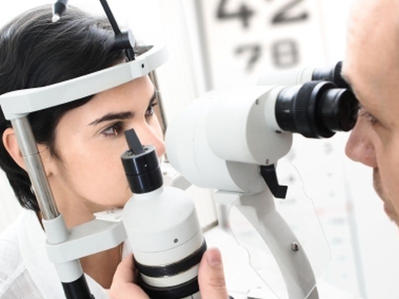
Ophthalmoscopic examination reveals engorged and tortuous retinal veins, a hyperemic and swollen optic disk (optic nerve head), and retinal hemorrhages around the disk but not into the retinal periphery. Isolated disk edema (eg, caused by optic neuritis or ischemic optic neuropathy) without the retinal findings indicative of elevated CSF pressure is not considered papilledema.

Image provided by James Garrity, MD.
In the early stages, visual acuity and pupillary response to light are usually normal and become abnormal only after the condition is well advanced. Visual field testing may detect an enlarged blind spot. Later, visual field testing may show defects typical of nerve fiber bundle defects and loss of peripheral vision.
Diagnosis
Clinical evaluation
Immediate neuroimaging
The degree of disk swelling can be quantified by comparing the plus lens numbers needed to focus an ophthalmoscope on the most elevated portion of the disk and on the unaffected portion of the retina. Swelling can also be quantified by measuring nerve fiber layer thickness using optical coherence tomography (OCT); OCT is done to quantify the degree of papilledema so that changes can be monitored.
Differentiating papilledema from other causes of a swollen optic disk, such as optic neuritis, ischemic optic neuropathy, hypotony, central retinal vein occlusion, uveitis, or pseudo swollen disks (eg, optic nerve drusen), requires a thorough ophthalmologic evaluation. If papilledema is suspected clinically, MRI with gadolinium contrast or CT with contrast is done immediately to exclude causes such as an intracranial mass. Lumbar puncture (seeLumbar puncture (spinal tap)) with measurement of CSF pressure and analysis of CSF should be done if a mass lesion has been ruled out. Lumbar puncture in patients with intracranial mass lesions can result in brain stem herniation. B-scan ultrasonography is the best diagnostic tool for the pseudo disk edema of optic nerve drusen.
Treatment
Treatment of underlying disorder
Urgent treatment of the underlying disorder is indicated to decrease intracranial pressure. If intracranial pressure is not reduced, secondary optic nerve atrophy and vision loss eventually occur, along with other serious neurologic sequelae.
Key Points
Papilledema indicates increased intracranial pressure.
In addition to bilateral hyperemic and swollen optic disks (optic nerve heads), patients typically have engorged and tortuous retinal veins, and retinal hemorrhages around the disk but not into the retinal periphery.
Funduscopic abnormalities usually precede visual disturbances.
Do immediate neuroimaging and, if no mass lesion is seen, obtain CSF for analysis and measure CSF pressure with a lumbar puncture.
Treat the underlying disorder.
Last full review/revision April 2014 by James Garrity, MD
Resources In This Article
Optic Nerve DisordersThe Optic PathwayHereditary Optic NeuropathiesIschemic Optic NeuropathyOptic Neuritis
Papilledema
* THIS IS THE PROFESSIONAL VERSION. *
CONSUMERS:Tap here for the
Go to Consumer Version
Also of Interest
NEWSHealthDay
January 2016 Briefing - Ophthalmology
Here are what the editors at HealthDay consider to be the most important developments in Ophthalmology for January 2016. This roundup includes the latest research news from journal articles, as well as the FDA approvals and regulatory changes that are the most likely to affect clinical practice. ...
![]()
TEST YOURKnowledge
Which of the following disorders is most likely a contributing factor in patients with floaters?
DiabetesCataractsDry eyePresbyopiaAM I CORRECT?

Glaucoma
Inside the eye are two fluid-filled chambers. Intraocular fluid, or eye fluid, is responsible for maintaining the correct amount of pressure inside the eye, which keeps the shape of the eye intact. This fluid is clear and watery in the front, or anterior chamber of the eye. The back, or posterior...

NEWSHealthDay
Nitrate Intake Linked to Primary Open-Angle Glaucoma Risk
FRIDAY, Jan. 15, 2016 (HealthDay News) -- Higher dietary nitrate and green leafy vegetable intake is associated with reduced risk of primary open-angle glaucoma (POAG), according to a study published online Jan. 14 in JAMA Ophthalmology . Jae H. Kang, Sc.D., from Brigham & Women's Hospital...
NEWSHealthDay
Earlier AMD Onset With Rare Genetic Variants
MONDAY, Jan. 18, 2016 (HealthDay News) -- Patients with age-related macular degeneration (AMD) carrying rare variants have earlier age at symptom onset and a higher prevalence of positive family history than noncarriers, according to a study published online Jan. 14 in JAMA Ophthalmology . Nicole...

NEWSHealthDay
Yoga Positions Linked to Increase in Intraocular Pressure
WEDNESDAY, Jan. 13, 2016 (HealthDay News) -- For individuals with and without open-angle glaucoma, intraocular pressure (IOP) increases shortly after starting yoga exercises with head-down positions, according to a study published online Dec. 23 in PLOS ONE . Jessica V. Jasien, from the New York...
NEWSHealthDay
January 2016 Briefing - Ophthalmology
Here are what the editors at HealthDay consider to be the most important developments in Ophthalmology for January 2016. This roundup includes the latest research news from journal articles, as well as the FDA approvals and regulatory changes that are the most likely to affect clinical practice. ...
![]()
TEST YOURKnowledge
Which of the following disorders is most likely a contributing factor in patients with floaters?
DiabetesCataractsDry eyePresbyopiaAM I CORRECT?
PreviousNext123456
![]()
MSD and the MSD Manuals
Merck & Co., Inc., Kenilworth, NJ, USA (known as MSD outside of the US and Canada) is a global healthcare leader working to help the world be well. From developing new therapies that treat and prevent disease to helping people in need, we are committed to improving health and well-being around the world. The Manual was first published in 1899 as a service to the community. The legacy of this great resource continues as the Merck Manual in the US and Canada and the MSD Manual outside of North America. Learn more about our commitment to Global Medical Knowledge.
ABOUTPERMISSIONSPRIVACYDISCLAIMERCONTRIBUTORSTERMS OF USELICENSINGCONTACT USGLOBAL MEDICAL KNOWLEDGE
© 2015 Merck Sharp & Dohme Corp., a subsidiary of Merck & Co., Inc., Kenilworth, NJ, USA
No comments:
Post a Comment